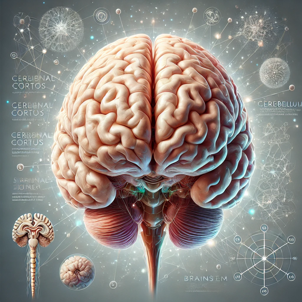
Let’s now discuss Neuroanatomy.
Neuroanatomy: Key Aspects
Neuroanatomy is the study of the structure and organization of the nervous system. It focuses on understanding how different parts of the brain, spinal cord, and peripheral nervous system are organized and how they interconnect to support various bodily functions and behaviors. Neuroanatomy seeks to map out the intricate networks of neurons that make up the nervous system, studying both the macroscopic level (brain regions, nerves, and pathways) and the microscopic level (neuronal connections and synapses).
This field is critical for understanding how different regions of the brain control specific behaviors, processes, and bodily functions. For example, the cerebral cortex is responsible for higher cognitive functions like thinking and decision-making, while the cerebellum plays a key role in motor coordination and balance. Neuroanatomists also examine the brain’s organization in relation to sensory processing, motor control, emotions, and memory, providing the anatomical basis for other fields of neuroscience to investigate how these structures function and interact.
Impact on People
Neuroanatomy is foundational for both basic science and clinical applications. Understanding the precise organization of the nervous system is essential for diagnosing and treating neurological conditions, such as strokes, brain injuries, tumors, and degenerative diseases like multiple sclerosis or ALS (Amyotrophic Lateral Sclerosis). Neuroanatomy also plays a critical role in neurosurgery, where detailed knowledge of the brain’s structure helps surgeons avoid damaging critical areas during operations.
For the general population, advances in neuroanatomy can lead to improved surgical techniques, better diagnostic imaging tools (such as MRI and CT scans), and a deeper understanding of how disorders affect brain function. For example, neuroanatomical research has allowed doctors to pinpoint regions affected by Alzheimer’s disease or to understand the neural circuits involved in Parkinson’s disease, leading to more targeted treatments.
A Day in the Life of a Neuroanatomist
Neuroanatomists often work in academic or clinical settings, where they spend time studying both human and animal brains. A typical day might include a combination of laboratory work, imaging studies, and collaboration with other researchers. Here’s a glimpse of what their daily activities might involve:
-
Morning: Brain Dissections or Imaging Studies
The day might begin with dissecting brain tissues to better understand their structure and organization. This could involve studying post-mortem human brains or animal models to identify how different brain regions are connected. Alternatively, they might analyze brain scans using tools like MRI or DTI (Diffusion Tensor Imaging) to map neural pathways in living subjects. -
Midday: Data Collection and Mapping
After dissections or imaging studies, neuroanatomists spend time collecting data on the brain’s structure. This might involve using computer software to create 3D reconstructions of brain pathways or using specialized techniques to trace neural connections between brain regions. They may also study stained brain slices under a microscope to examine individual neurons and their synapses. -
Afternoon: Research Meetings and Collaboration
Collaboration is key in neuroscience, and neuroanatomists often meet with other neuroscientists, neurosurgeons, or radiologists to discuss their findings and plan joint studies. They might also collaborate with specialists in other fields, such as cognitive neuroscience, to link anatomical data with functional insights. Some neuroanatomists work closely with clinicians to apply their research to real-world medical problems. -
Evening: Writing and Presentation Preparation
Neuroanatomists spend time writing research papers or preparing presentations to share their findings at conferences or in academic journals. They might also be involved in teaching students about neuroanatomy, either through lectures or hands-on dissections in a medical or university setting.
Skills and Knowledge Needed for Success
Success in neuroanatomy requires a combination of theoretical knowledge, technical expertise, and analytical skills. Here’s what it takes to excel in this field:
-
Strong Background in Anatomy and Neuroscience
Neuroanatomists must have a detailed understanding of general anatomy and the nervous system. This includes knowledge of different brain regions, neural pathways, and the functions associated with various structures. They need to be familiar with both macroscopic structures (like lobes and tracts) and microscopic structures (like neurons and synapses). -
Imaging and Dissection Skills
Neuroanatomy often requires the use of advanced imaging techniques like MRI, CT scans, and DTI to visualize the brain. Neuroanatomists must be proficient in analyzing these images and understanding what they reveal about brain structure. Additionally, they need to be skilled in dissection techniques, whether they are working with animal models or post-mortem human brains, to accurately observe and document anatomical features. -
Data Analysis and 3D Mapping
Mapping the brain requires the ability to interpret complex data and create detailed diagrams or 3D models of brain structures. Neuroanatomists often use specialized software to trace neural pathways, reconstruct brain regions, or analyze the connectivity between different parts of the brain. This requires strong analytical and technical skills, as well as the ability to work with imaging data. -
Attention to Detail and Precision
Neuroanatomy is highly detailed work. Neuroanatomists must be meticulous when documenting findings, whether through imaging, dissection, or microscopic examination. Small mistakes can lead to incorrect conclusions about the brain’s organization, so precision is critical. -
Collaboration and Communication
Because neuroanatomy overlaps with other fields, such as neurosurgery and radiology, effective collaboration is essential. Neuroanatomists need to communicate their findings clearly to colleagues from different disciplines, often contributing to multidisciplinary research projects. They must also be capable of teaching complex concepts to students or presenting their research at scientific conferences. -
Problem-Solving and Critical Thinking
Neuroanatomy often involves solving complex puzzles about how different brain regions are connected and how these connections contribute to behavior or disease. Neuroanatomists need to think critically about their findings and approach problems creatively to develop new insights into brain structure and function.
Academic Pathway
A career in neuroanatomy typically starts with a bachelor’s degree in neuroscience, biology, anatomy, or a related field, followed by a graduate program specializing in neuroanatomy or a closely related discipline. Ph.D. programs are common for those interested in research, though medical degrees are also an option for neuroanatomists working in clinical settings. Training in advanced imaging techniques and neuroscience research methods is also essential. Postdoctoral work or specialized training in neuroimaging or brain mapping techniques may be required for those aiming to lead their own research projects or work in academia.
Neuroanatomy is a crucial field that bridges our understanding of brain structure with brain function, helping researchers and clinicians diagnose and treat neurological conditions more effectively. It provides the foundational map of the nervous system that other fields of neuroscience rely on for their work.
